


Table of contents
Hemangiomas are benign non-cancerous tumors found on blood vessels that occur in around 10% of people worldwide, most often without showing any noticeable symptoms.
Hemangiomas are usually incidental findings on imaging tests and do not require treatment, though regular monitoring may be beneficial; symptoms may include localized pain and the corduroy sign (a pattern of thickened bone bands that resemble stripes on corduroy fabric), although more serious consequences could include spinal cord compression.

What Causes Hemangiomas of the Spine?
The exact cause of spinal hemangiomas remains unknown. These noncancerous tumors consist of abnormal blood vessels and stand as the most frequent primary tumors in the spine.
These vascular lesions generally arise from normal capillary and venous structures and are often situated in the upper-to-mid thoracic or lower lumbar spine.
Hemangiomas are prevalent, affecting around 10% of the global population. In most instances, no symptoms emerge; only a small portion of these lesions generate symptoms, mainly back pain and neurologic issues.
The risk of bone hemangiomas is elevated in women and older individuals, especially beyond 50 years of age. If identified during other imaging and devoid of symptoms, treatment may be unnecessary.

Potential Causes
Hemangiomas are noncancerous tumors composed of abnormal blood vessels that can appear anywhere on the body, from soft tissues and bones, to soft and hard tissues like skin. Bone hemangiomas, or vertebral hemangiomas or VHs, tend to occur more commonly in thoracic and lumbar sections of spine; most are often asymptomatic and do not need treatment.
Scholl underwent an MRI which confirmed his suspicions of spinal hemangioma in his seventh thoracic vertebra, situated near his middle back region. This growth was pressing on nerves within his spinal cord and causing severe discomfort and numbness throughout his arms, legs, and feet.
Note that Hemangiomas in Bone can be difficult to diagnose accurately and timely. They appear as honeycomb-shaped masses within bones, often mistakingn for arteriovenous malformations (AVMs), hemangiomas in internal organs or even Venous Veno-Occlusive Diseases (VVODs).
Their symptoms resemble those of many spinal disorders making timely diagnosis essential in providing timely treatments.
Risk Factors
Hemangiomas are non-cancerous tumors consisting of abnormal blood vessels that are extremely common. While they can appear anywhere on the body, they seem more frequent as people age. Although non-cancerous, hemangiomas may still cause problems by compressing nerves or the spinal canal and potentially becoming cancerous over time.
Vertebral hemangiomas (often known as vertebral hemangiomas) can be especially problematic. Though relatively uncommon, if left untreated promptly these spinal lesions can lead to pain, weakness and loss of bladder/bowel control.
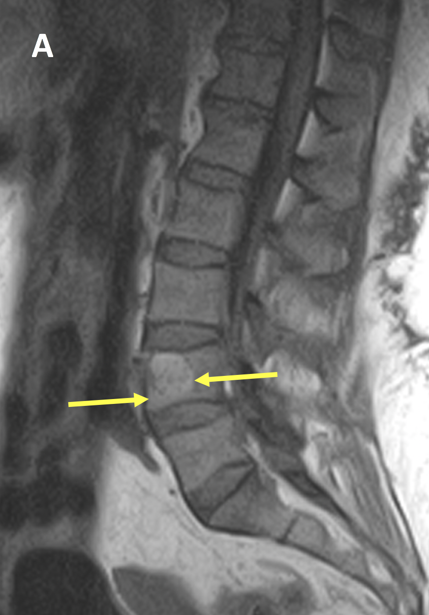
Hemangiomas of the spine are typically diagnosed through an X-ray and CT scans, or occasionally an MRI.
Hemangiomas are easily recognized on radiographs as having honeycomb or lattice-like structures; CT and MRI scans reveal them as dense bone areas with honeycomb patterns or lattice designs; they may even mimic malignant tumors; therefore some patients undergo additional testing in order to “rule out” more serious diagnoses.
Symptoms
Hemangiomas are benign blood vessel tumors found most commonly in the spine or elsewhere on the body. While not malignant (cancerous), they can expand and cause pain or spinal cord compression in rare instances.
Most hemangiomas remain asymptomatic until detected incidentally through imaging scans of the spine; in these instances however they should be managed immediately as symptoms may emerge as they expand further or grow larger than originally anticipated.
Spinal hemangiomas may be mistaken for other tumors visible through imaging tests, so an MRI or CT scan may be performed to check for their characteristic pattern of bone growth.
Hemangiomas in your spine can cause pain, numbness or tingling in arms and legs when they compress nerves or the spinal cord; they’re more likely to become painful as you age as well as located lower thoracic or lumbar vertebrae and more likely to manifest themselves symptomatically than ever.
Diagnosis
Hemangiomas are often discovered during CT or MRI exams performed for other reasons. Because hemangiomas usually have a distinctive pattern, these imaging tests make diagnosing them easy.
Hemangiomas in the spine are usually unnoticed and do not require treatment, however in more aggressive cases intervention may be needed to avoid complications such as pain, radiculopathy, spinal canal encroachment or compression of nerves.
Aggressive hemangiomas are more prevalent in the thoracic and lumbar spine regions, often being associated with radiologic features of osseous expansion, destruction of vertebral cortex destruction, and extension into posterior elements such as pedicles or epidural space.
Furthermore, aggressive hemangiomas often exhibit an “expanse-like” appearance on contrast MRI scans.
What Are the Symptoms of Spinal Hemangiomas

Spinal hemangiomas, common benign tumors from blood vessels, are the most frequent benign spinal tumors. Most cases are symptom-free. Yet, they can sometimes induce pain and neurological problems.
Symptoms of spinal hemangiomas often mirror those of other spine conditions, including:
- Back pain at the tumor site
- Radiating nerve pain due to pressure or inflammation from the tumor, causing discomfort
- Pain extending to arms or legs
- Arm or leg weakness, numbness, or clumsiness
- Impaired bowel or bladder control
Symptomatic hemangiomas comprise <1% of all cases and are more prevalent in women. If suspecting a hemangioma, seek proper diagnosis.
How Are Spinal Hemangiomas Diagnosed
Diagnosing spinal hemangiomas involves non-symptomatic discovery during imaging for unrelated issues, though they can cause discomfort.
Medical professionals diagnose them through X-rays, CT scans, and MRIs, identifying their size, location, and impact on the spinal cord and nerves.
A CT scan might display a bone’s polka dot appearance, signifying a hemangioma. If so, an MRI is ordered, revealing if the tumor affects the spinal cord or canal. MRIs use magnets, radio waves, and computers for precise spine images.
These hemangiomas are commonly found in upper-to-mid thoracic or lower lumbar spine regions. If incidentally discovered and symptom-free during unrelated imaging, treatment might be unnecessary.
What Are the Treatment Options for Spinal Hemangiomas
Spinal hemangiomas, benign tumors commonly found in mid-back (thoracic) and lower back (lumbar), are usually symptom-free. However, they can cause pain and neurological problems in some cases.
Treatment options depend on tumor size, location, and symptoms. Incidental, symptom-free findings during other imaging might not need treatment. But, if pain or neurological issues arise, options include:
- Embolization: Minimally invasive, it halts tumor blood flow.
- Ethanol injections: Shrinkage induced through alcohol injections.
- Kyphoplasty: Treats spine compression fractures caused by hemangioma.
- Resection: Surgical removal of tumor or affected vertebra.
- Radiation therapy: Eases hemangioma-induced pain.
- Laminectomy: Surgery to release spinal cord or nerve pressure.
- Instrumented fusion and fixation: Stabilizes vertebra with grafts, rods, and screws if needed.
Remember, most spinal hemangiomas don’t necessitate treatment; only symptomatic cases require it.


